Peripheral Vision and Visual Pathways
Objective:
To explain in short essays or diagrams how changes in peripheral vision can be associated with damage to the retina or central neural pathways, or reflect tumors in the brain, at the level of 85% proficiency for each student.
In order to achieve this objective, you will need to be able to:
- Measure peripheral vision in two axes.
- Explain the anatomy of the visual pathways
Materials:
Group Supplies:
colored pencils or pens
visual field diagrams
four colors of string to represent axons
four colors of beads to represent cell bodies
Lab Supplies:
disc perimetry device and target wand
Methods and Results:
Peripheral Vision
- To test the right eye, have the subject occlude the left eye.
- Hold the large plastic half disc horizontally against the nose to test the temporal and nasal fields.
- Have the subject look at the center dot.
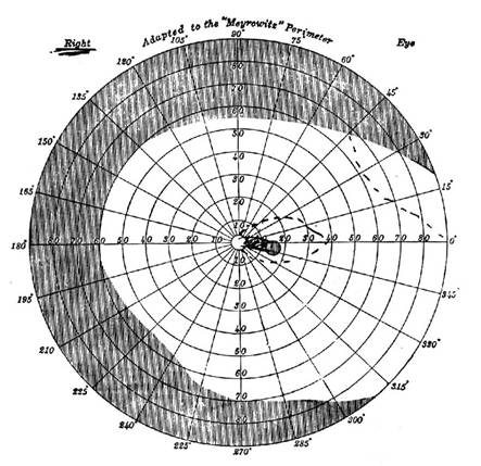
- To test the temporal field, hold the wand 1 cm to the temple side of the center dot and move outward until the peripheral target disappears. Repeat the test starting at 90o and move toward the center. Record the point of disappearance and appearance in degrees on the temporal side of the visual field diagram for the RIGHT eye.
- To test the nasal field, hold the wand near the center dot and move outward until the peripheral target disappears. Repeat the test starting at 90o and move toward the center. Record the point of disappearance and appearance in degrees on the nasal side of the visual field diagram for the RIGHT eye.
- Hold the large plastic half disc vertically against the nose to test the superior and inferior fields.
- To test the superior field, hold the wand near the center dot and move upward until the peripheral target disappears. Repeat the test starting at 90o and move downward to the center. Record the point of disappearance and appearance in degrees on the superior field of the visual field diagram for the RIGHT eye.
- To test the inferior field, hold the wand near the center dot and move downward until the peripheral target disappears. Repeat the test starting at 90o and move upward to the center. Record the point of disappearance and appearance in degrees on the inferior field of the visual field diagram for the RIGHT eye.
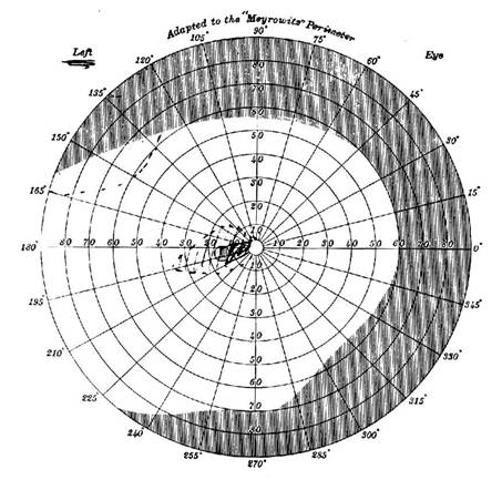
- Repeat for the LEFT eye with the right eye occluded.
Visual Field Diagram – RIGHT Eye
|
|
Nasal Field |
Temporal Field |
|
Superior Field
Inferior Field |
|
|
Visual Field Diagram – LEFT Eye
|
|
Nasal Field |
Temporal Field |
|
Superior Field
Inferior Field |
|
|
Visual Pathways
Construct the visual pathway using different colored beads and string. Include the cell bodies, axons, and synaptic sites for each pathway. Use beads to represent cell bodies and string to represent axons. Use a sheet of paper and label each nucleus and tract as they are relevant.
Use the following description, table and illustrations as a guide.
Visual pathways
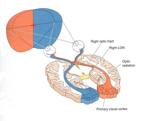
- Axons from the retinal ganglion cells travel through the optic nerves, optic chiasm, and optic tracts to reach the Lateral Geniculate Nucleus of the Thalamus
- Axons from the retinal ganglion cells of the left half of each retina synapse on neurons in the Left Lateral Geniculate Nucleus of the Thalamus.
- Axons from the retinal ganglion cells of the right half of each retina synapse on neurons in the Right Lateral Geniculate Nucleus of the Thalamus
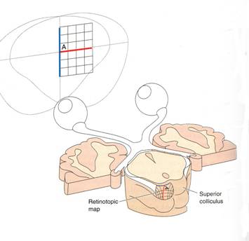
- Axons from the retinal ganglion cells also synapse on neurons in the Superior Colliculus which contain a sensory map and coordinates orientation of the eyes, head and neck toward visual stimuli.
- Neurons in the Left Lateral Geniculate Nucleus of the Thalamus synapse on neurons in the Left Primary Visual Cortex.
- Neurons in the Right Lateral Geniculate Nucleus of the Thalamus synapse on neurons in the Right Primary Visual Cortex.
- Both the Lateral Geniculate Nuclei and the Primary Visual Cortex contain a sensory map of the entire visual field and process (filter) visual signals.
Organization of the Visual Pathways
|
Left Eye |
Right Eye |
||
|
Left Half |
Right Half |
Left Half |
Right Half |
|
Left Optic Nerve |
Right Optic Nerve |
||
|
Optic Chiasm |
|||
|
Left Optic Tract |
Right Optic Tract |
Left Optic Tract |
Right Optic Tract |
|
Left Superior Colliculus Left LGN |
Right Superior Colliculus RightLGN |
Left Superior Colliculus Left LGN |
Right Superior Colliculus RightLGN |
|
Left Visual Cortex |
Right Visual Cortex |
Left Visual Cortex |
Right Visual Cortex |
Pathways to Primary Visual Cortex
|
|
Pathways to Superior Colliculus
|
|
Discussion:
- The retina extends farther forward on the medial (inside) of the eye than the lateral (outside). Did this affect the findings?
- Why is the superior field less than the inferior field?
- What accounts for the temporary target disappearance sometimes noted when testing the temporal fields?
- Could retinal damage be localized based on the results of this test?
- Think of the situation where reduced field "tunnel vision" could be dangerous.
- Reduced field of vision is often a symptom of glaucoma. Can you offer an explanation for this occurrence?
- Diagram the pathway and neurons involved from the retina to the visual cortex.
- Describe how the tectal system (superior colliculus) pathways are involved in the control of the extrinsic eye muscles, circular muscle fibers of the iris, and ciliary muscle.
- Describe the visual disturbance that often accompanies a pituitary tumor. Explain why this particular pattern is observed.
- If a patient reported that he could not see with his left eye, how could you tell if the problem was the optic nerve or the optic radiation? Explain.
© David G. Ward, Ph.D. Last modified by wardd 23 May, 2006