Somatoensory and Pain Pathways
Receptor Density and Somatosensory Cortex
Human skin contains several different sense receptors that respond to mechanical and thermal stimuli (e.g., touch, pressure, pain, cold, and heat). These receptors help us explore and determine the characteristics of our external environment. See Figure 1.
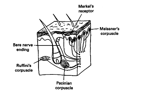
Figure 1. Types of sense receptors in the skin.
A sense receptor is a specialized cell that transduces (converts) a physical or chemical stimulus into action potentials. These action potentials produced by the receptors conduct to the spinal cord and brain for processing and interpretation. The message that is sent to the central nervous system (CNS) is always a train of action potentials, regardless of the kind of stimulus that excites a particular receptor. Sensory receptors that respond to touch send action potentials through axons that enter the dorsal columns of the spinal cord and ascend to the medulla of the brainstem. These axons then make connections (synapses) with another pathway within the medulla. It is here that the pathway crosses over the brain midline and then continues to the thalamus. The final pathway begins in the thalamus and continues to the specific region of the sensory cortex, as shown in Figure 2. This pathway effectively gives one side of the brain responsibility for the opposite side of the body. This phenomenon is observed when a head injury or disease of the right side of the brain leads to physiological problems in a patient's left side.
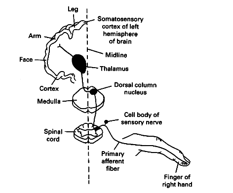
Figure 2. The dorsal column pathway for carrying somatosensory signals.
Most of the information about touch centers in a thin, convoluted surface layer of the cerebrum of the human brain called the somatosensory cortex. Each point on this band of sensory cortex contains densely packed cells that correspond to sensory receptors from different parts of the body, as shown in Figure 3. The specific amount of space on the somatosensory cortex of the brain that is dedicated to sensing each body part is proportional to the density of the sensory receptors in that particular body region. For example, relatively few receptors are located in the upper arm; therefore, the upper arm area in the somatosensory cortex is small. In contrast, the density of receptors in the lips is very high, so the lip area of the cortex is large. The two-point discrimination method described in this activity approximates a map of the entire body as "sensed" by the cortex. The "picture" of the body on the somatosensory cortex is the "homunculus," which means "little person."
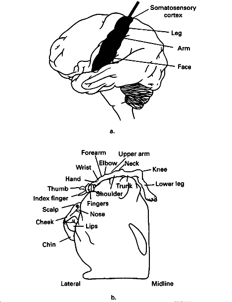
Figure 3. The somatotopic mapping of the somatosensory cortex.
The two-point discrimination method determines the minimum distance sensed between two points of touch. This technique determines the approximate size of the receptive field that senses light touch. In this procedure, the two points of a caliper lightly touch the skin at the same time and the subject determines whether one or two points of the stimulus are felt. Two points placed on the tips of the fingers can be distinguished as two separate points even when the points are as close together as two millimeters. In contrast, on the subject's back the points must be about 40 millimeters apart before they are sensed as two separate stimuli (Martin & Jessell, 1991, p. 346). The reason the discrimination of the finger is better than the back is that the finger has a much larger number of specialized touch receptors than the back or other regions of the body.
In this activity, students will locate touch receptors on the skin and estimate their relative numbers in different parts of the body. By calculating the reciprocals of the two-point discrimination numbers from the skin, students will be able to predict the relative size of the region of the sensory cortex responsible for detecting sensation in a given area of skin. They can then consider how the resulting pattern may affect human life and evolution.
Touch sensors are found throughout a person's body. Students may wonder why they are not normally aware of the touch caused by everyday stimuli such as their clothes and sitting in a chair. These touch sensors are designed to be excited for a short period of time. After a while they do not respond any more to the constant stimulation; in other words, they adapt to a stimulus. If the touch is changed slightly by movement or touch shifts, the sensors again become responsive for a short period of time.
Regions in the brain corresponding to touch receptors on the skin can be reconfigured when the area of the body they are responsible for is missing, as in amputation. The brain may remodel the sensory neurons so that they are used for another function.
Pain Pathways:
Pain is very important to our survival. Although pain is defined as the perception of a noxious (i.e., tissue damaging) stimulus, pain can also occur in the absence of injury or long after an injury has healed. Pain provides humans with information about tissue-damaging stimuli, and thus enables them to protect themselves from greater damage. Pain is protective in two ways. First, it removes a person from stimuli that cause tissue damage through withdrawal reflexes. Second, learning associated with pain causes the person to avoid stimuli that previously caused pain. In humans, pain often initiates the search for medical assistance and helps us to pinpoint the underlying cause of disease. In addition, pain protects sensitive body regions from further injury and thus facilitates recovery from injury.
Specialized sensory neurons called nociceptors transmit messages about tissue damage from the skin or other site of injury to the spinal cord. See Figure 1. Nociceptors respond exclusively to stimuli that damage the skin and other tissues.
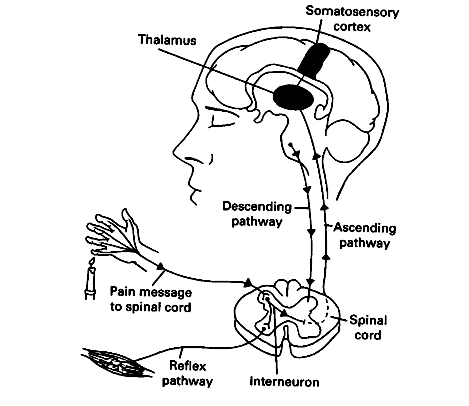
Figure 4. The spinal and spinothalamic pathways for carrying pain signals.
Two things happen once the message reaches the spinal cord. Spinal interneurons transmit the message to motor neurons that synapse with muscles involved in withdrawal reflexes. This reflex circuit removes the injured limb from the stimulus. Simultaneously, a message travels to the thalamus, which relays the message to the somatosensory cortex. Because of the difference in distance in these two pathways, nociceptive reflexes occur before pain messages reach the brain. Although much is known about the pathways from injured tissue to the brain, the interpretation of pain in the brain is not completely understood (Guyton, 1991).
Several types of neurons carry information about painful stimuli to the spinal cord (Martin & Jessell, 1991, pp. 349-351). When students pinch the finger web, they experience two types of pain, as shown in Figure 5. It is hypothesized that different types of nociceptors carry these two types of pain. Small, myelinated fibers (A delta fibers) appear to carry sharp, pricking sensations, whereas unmyelinated fibers (C fibers) appear to carry a diffuse, throbbing pain. A delta fibers carry messages at a velocity of 6 to 30 m/second and C fibers at a velocity of 0.5 to 2.0 m/second (Guyton, 1991, p. 522). The long- lasting throbbing pain is probably the result of prolonged activity in C fibers. Students who have hit a finger with a hammer or dropped something on a toe have probably experienced these two types of pain.
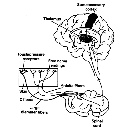
Figure 5. Type of pain and pain fibers.
What is life like for a person who cannot feel pain? The protective value of pain is demonstrated easily by examining individuals who are insensitive to pain. People with a rare condition called congenital insensitivity to pain lack the neural apparatus for detecting harmful stimuli. They repeatedly injure themselves because they do not avoid hot objects, intense pressure, extreme twisting, or corrosive substances, and, thus, end up with burns, pressure sores, missing digits, and damaged joints. Specific examples of some of these injuries are:
- The little girl who poked a pencil through her cheek.
- A baby girl who bit off the tip of one of her fingers and watched it bleed.
- A young woman who died of spine damage because she did not receive the normal discomfort signals from her joints telling her to change her posture--she never moved in her sleep, for ex- ample (Fields, 1987).
Pain is an unpleasant sensory experience associated with actual or potential damage to the body, or perception of such damage. It is a subjective experience. The perception of pain varies with the intensity of the stimulus and the affective or emotional state of the individual. Memories of events associated with extreme pain persist for a long time. This persistence should become evident as you look at the ages listed when their worst pain occurred.
Mental state is known to have a powerful influence over pain. For example, an athlete may not notice a twisted ankle until after the competition is over. Similarly, soldiers in battle often continue to fight even after sustaining serious injury, and they may report afterwards that they experienced no pain until after battle. The scientific explanation for this phenomenon is that the brain not only receives pain messages, but also has a descending system of neurons that suppresses pain messages, as shown in Figure 4. This system inhibits cells in the spinal cord that transmit pain signals. The descending system is thus a pathway for natural pain modulation. Opioids that occur naturally such as the endorphins are important neurotransmitters in some of these descending pathways (Carola, Harley & Noback, 1990, p. 432). Endorphins appear to be released by the brain in times of stress (e.g., attack from a predator) in order to minimize pain that may detract from an organism's ability to escape. Extreme exercise may also cause the release of endorphins; leading to the "natural high" experienced by serious runners (Carola, Harley & Noback, 1990, p. 432).
Physicians make use of these endogenous pain modulator systems by treating severe pain with opioids derived from plants such as morphine or codeine (Society for Neuroscience, 1990, pp. 19-20). These drugs bind to endogenous opioid receptors in the central nervous system (CNS) to relieve pain much as naturally occurring opioids do. See Figure 6. Physicians can also treat pain by blocking nerve conduction with anesthetics or by surgically cutting a nerve. Cutting a nerve causes a permanent loss of all sensations carried by the cut nerve, and, yet, surgery often does not alleviate persistent pain problems.
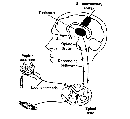
Figure 6. Drugs that affect pain pathways.
In contrast, simply simply rubbing the injury, applying ice, or taking aspirin or other over-the-counter painkillers often reduces mild pains. These mechanisms work by reducing neural transmission either indirectly via inhibiting inflammation (e.g., aspirin, cold water) or via interfering with nociceptive messages in the spinal cord (e.g., rubbing the skin activates touch fibers that inhibit nociceptive neurons in the spinal cord).
© David G. Ward, Ph.D. Last modified by wardd 23 May, 2006