Spinal and StretchReflexes
A reflex is an involuntary neural response to a specific sensory stimulus that threatens the survival or homeostatic state of an organism. Reflexes exist in the most primitive of species, usually with a protective function for animals when they encounter external and internal stimuli. A primitive example of this protective reflex is the gill withdrawal reflex of the sea slug Aplysia. In humans and other vertebrates, protective reflexes have been maintained and expanded in number. Examples are the gag reflex that occurs when objects touch the sides or the back of the throat, and the carotid sinus reflex that restores blood pressure to normal when baroreceptors detect an increase in blood pressure. A second type of reflex, the stretch reflex, has evolved to help maintain muscle tone that is important for posture and movement.
An understanding of the neural circuitry underlying each type of reflex also has clinical significance. When physicians test for potential damage to various components of the nervous system, they often begin by attempting to elicit reflex responses to the appropriate stimuli. Examples of reflexes with protective and diagnostic importance are the flexor withdrawal reflex, the corneal (blink) reflex, and the accommodation reflex. The loss of the pupillary light reflex in a comatose patient, described under the section Protective reflex, often indicates extreme damage deep in the brain. Loss of the knee-jerk response may indicate problems with muscle tone, chronic diabetes, or damage at the L2, L3, or L4 vertebral level.
For each reflex resulting in a specific motor output, the neural pathway linking the sensory receptor to the central nervous system (CNS) may be studied. In this activity, components of the neural circuitry of reflexes will be studied. These components include a sensory receptor, a sensory (afferent) neuron, a reflex center in the CNS that may involve one or more levels of integration, a motor (efferent) neuron, and a muscle (efferent). See Figure 1.
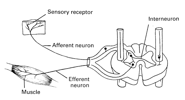
Figure 1. Components of neural circuitry of reflexes.
Stretch Reflex
The knee-jerk (patellar) reflex is a stretch reflex. See Figure 2. Tapping the patellar tendon stretches striated muscle fibers in the quadriceps, resulting in activation of specialized sensory receptors within the muscle called muscle spindles. Sensory neurons connected to the muscle spindles are activated and carry the stimulus to the spinal cord, where they synapse onto a motor neuron. These motor neurons are triggered by a neurotransmitter and release a neurotransmitter back at the quadriceps muscle in a synaptic area of the striated muscle membrane called the neuromuscular junction. This results in shortening the length of the muscle fibers and knee extension. The knee-jerk and other tendon reflexes differ significantly from other types of reflexes in that no central nervous system interneurons are involved in the knee-jerk reflex. Under normal circumstances, brain pathways descend onto the same motor neurons, constantly modulating the strength of stretch reflexes. This modulation of muscle tone keeps muscles from being damaged during everyday movements. This process occurs not only in the quadriceps muscle, but also in other areas of the body as well. If students experience mild discomfort while participating in this activity, it is important to emphasize that this discomfort was felt only after the reflex response. Pain perception is processed by the brain and cannot be experienced until the message has had enough time to travel to the brain.
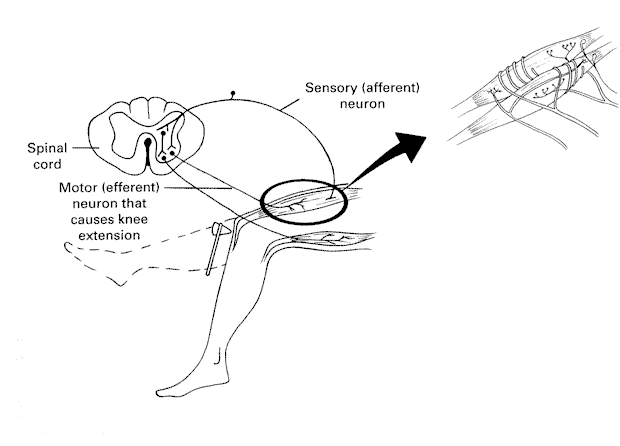
Figure 2. Stretch reflex.
© David G. Ward, Ph.D. Last modified by wardd 23 May, 2006